Command Palette
Search for a command to run...
Saying Goodbye to Manual Fluorescence Counting: UK Team Develops AI Workflow for Rabies Vaccine Evaluation With AUC of 0.999
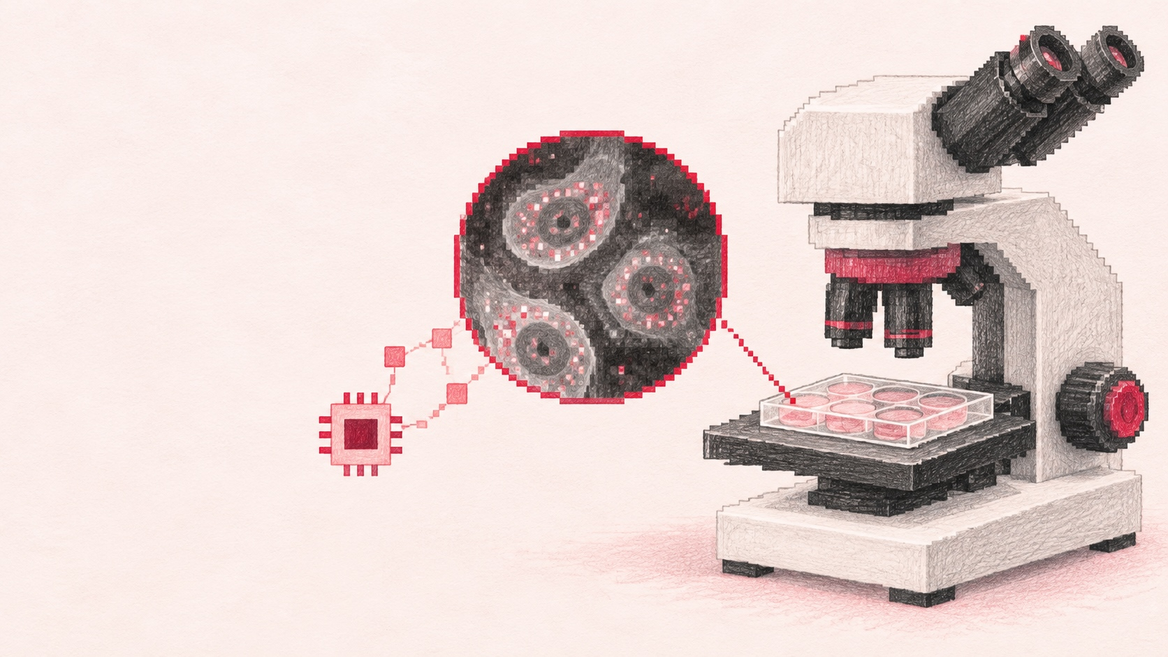
Rabies is a severe zoonotic disease that continues to cause tens of thousands of deaths annually in endemic regions. Although vaccination can effectively prevent rabies, the diagnosis of viral infection, assessment of immune response after vaccination, and related serological surveillance still rely on reliable laboratory testing methods. In cell culture testing, researchers typically need to rely on fluorescence microscopy imaging to determine whether cells are infected with rabies virus (RABV). The entire process involves extensive manual observation and interpretation of images, which is not only time-consuming but may also be affected by operator experience as well as factors such as weak fluorescence and imaging artifacts.
In recent years, advances in deep learning (DL) technology have significantly promoted the development of biomedical image analysis, including the assessment of viral infections. However, no previous studies have applied deep learning methods to the systematic fluorescence imaging analysis of rabies virus antigens. Since rabies virus typically does not cause obvious cytopathic effects or syncytium formation, the cellular morphological features relied upon by many AI-based methods have limited applicability in such detection.
Against this background, a research team from the University of Surrey in the UK proposed the first AI-based workflow for detecting rabies virus infection in BHK-21 cell cultures and quantifying antibody titers. The researchers selected three advanced deep learning architectures—ResNet, Swin Transformer V2, and MambaOut—and trained them using a large-scale dataset. In evaluations at three levels—image regions, single images, and wells—the MambaOut model consistently outperformed the other models.
At the well level, the model achieved an accuracy of 0.996, an F1 score of 0.993, and an area under the receiver operating characteristic curve (AUC-ROC) of 0.999. To assess the practical applicability of this method, researchers further tested the model using unseen images from the fluorescent antibody virus neutralization (FAVN) test recommended by the World Health Organization (WHO) and the World Organisation for Animal Health (WOAH). MambaOut showed complete consistency with ground truth annotations in predicting antibody concentrations, confirming the reliability of this workflow in vaccine evaluation and pet travel-related testing.
The relevant research results, titled "Deep learning enables rabies virus detection and antibody quantification in cell culture," have been published in Scientific Reports.
Research Highlights:
-
Constructed the first large-scale rabies virus imaging dataset
-
Achieved automation of high-throughput image analysis using AI, providing diagnostic laboratories with a scalable, time-saving solution, particularly suitable for countries and regions where rabies is endemic
-
The research results lay the foundation for AI-assisted rabies diagnosis and open new possibilities for applying deep learning technologies to other cell culture-based viral detection methods
Paper Link:
https://www.nature.com/articles/s41598-026-57955-8
Constructing the LyssaScan Dataset: Identifying Infection Signals from Fluorescence Images
The researchers first constructed the image dataset LyssaScan for RABV infection recognition, which covers various infection levels, staining reagents, and imaging conditions, simultaneously reflecting both biological differences and technical variations.
Specifically, researchers used BHK-21 cells infected with the standard challenge strain of rabies virus (RABV) 11 (CVS-11) to establish a standardized fluorescence microscopy imaging workflow. Cell culture was performed in flat-bottom, clear-bottom, black-walled 96-well plates. After 48 hours of culture, researchers fixed the cells and stained them with fluorescein isothiocyanate (FITC)-labeled polyclonal anti-rabies virus nucleocapsid antibodies (Fujirebio). Negative controls included wells with uninfected cells and wells containing only medium. Some cell cultures were stained with another FITC-conjugated antibody (Sifin). The complete imaging workflow is shown in the figure below:
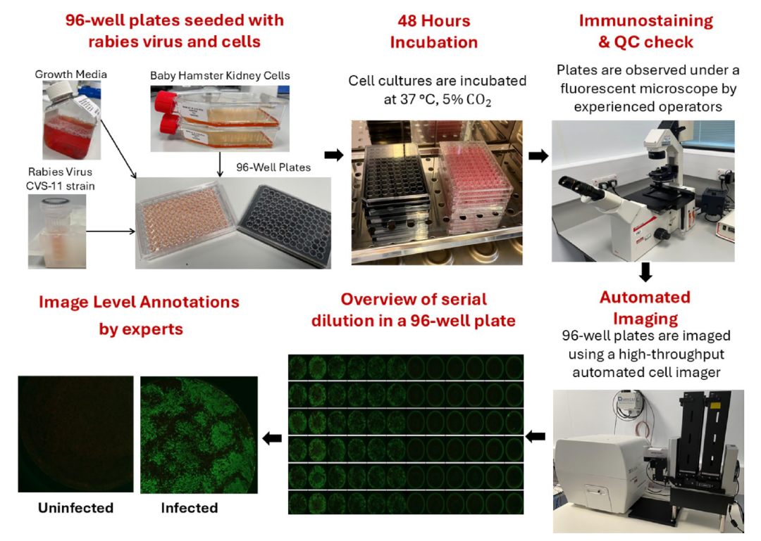
Automated workflow for dataset generation
Automated imaging was performed using the Cytation 5 cell imaging analyzer from Agilent Technologies, equipped with an objective lens whose model information is anomalously displayed in the original text, and an integrated wide-field camera. Researchers used GFP and Texas Red filter blocks to collect fluorescence signals generated by viral nucleocapsid staining and images for observing the cell monolayer, respectively. Each well was imaged at four corner positions, thereby generating four composite PNG images per well. Ultimately, a total of 2,344 composite images were collected for subsequent processing.
Subsequently, the images were resized to specific dimensions to allow for precise division into 16 non-overlapping image regions (patches), which were manually annotated by professionals to generate reliable ground truth labels. Cases where consensus could not be reached were marked as "ambiguous" and excluded from model development. In the end, the LyssaScan dataset contained a total of 36,822 image regions, with 9,750 infected samples and 27,072 uninfected samples.
Architectural Differences Among Three Models: From Convolutional Networks to Gated Convolutions
To compare the performance of different deep learning architectures in fluorescence image recognition tasks, researchers selected three models: ResNet-101, Swin Transformer V2 Small (SwinT-V2), and MambaOut Small. The parameter counts for these models are approximately 44.5 million, 47 million, and 48.5 million, respectively.
ResNet-101: A Convolutional Neural Network Based on Residual Connections
ResNet serves as the baseline model in this study. Its core feature is the introduction of skip connections, which improve gradient propagation as the network depth increases. By extracting image features layer by layer, ResNet can identify local fluorescence structures. However, its ability to discriminate complex backgrounds and faint infection signals may still be limited by its local feature representation capabilities.
SwinT-V2: Leveraging Self-Attention to Integrate Contextual Information
SwinT-V2 is a hierarchical vision Transformer that employs a shifted window self-attention mechanism. It integrates broader contextual information while extracting local features. For fluorescence images, infection foci may be small and can be mixed with background signals, staining variations, or other structures. Therefore, comprehensively considering both local appearances and surrounding environments helps enhance recognition capabilities.
MambaOut: Dynamically Regulating Features Through Gating Mechanisms
MambaOut in this study uses stacked gated CNN modules without state space model components. The gating mechanism dynamically regulates feature propagation, selectively enhancing valuable signals and suppressing background noise. This characteristic may be more suitable for identifying faint fluorescence foci embedded in complex backgrounds, which is one of the reasons researchers focused on its performance.
The three architectures represent technological approaches with complementary characteristics—Convolutional Neural Networks (CNN), Vision Transformers (ViT), and Gated Convolutional Neural Networks—providing a basis for systematically comparing different architectures.
Model training employed standard data augmentation methods, including random flipping, rotation, and adjustments to brightness and contrast, to improve model robustness. Models were initialized with ImageNet-1k pre-trained weights and fine-tuned on the LyssaScan dataset. Training was conducted on NVIDIA GeForce RTX 2080 Ti GPUs for 20 epochs with a batch size of 128.
The optimization process used the Adam optimizer. The formula for the initial learning rate was displayed abnormally in the original text and could not be accurately reconstructed, and a cosine annealing learning rate schedule was used. To address class imbalance, the study employed a weighted cross-entropy loss function, where class weights were inversely proportional to the sample frequency of each class.
During training (as shown in the figure below), features extracted by each model were first aggregated into a D-dimensional feature descriptor vector via Global Average Pooling (GAP), then input to a fully connected layer (FC) to generate logits, and finally optimized using the weighted cross-entropy loss function.
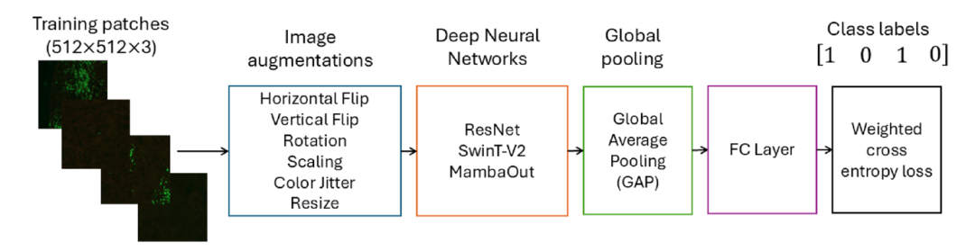
During the model training process
During the inference phase (as shown in the figure below), input image regions were resized and processed by the trained model. Extracted features were aggregated via Global Average Pooling, then passed through a fully connected layer and a Softmax layer to obtain predicted probabilities for each class. Subsequently, these probabilities were used to calculate various evaluation metrics for the models.
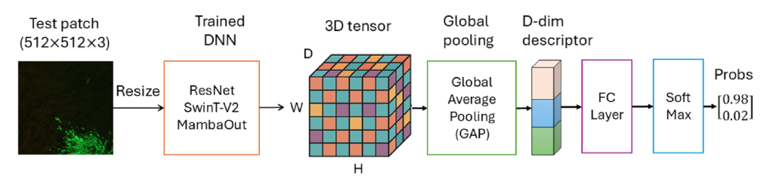
Model inference process
MambaOut Model Balances Recognition Accuracy and Antibody Quantification Capability
Researchers systematically evaluated the ResNet, Swin Transformer V2, and MambaOut models, comparing the performance of current advanced deep learning models in rabies virus detection tasks, providing a basis for the selection and application of relevant models.
Model Performance Evaluation
Model performance was evaluated at three levels. First, detection performance was assessed at the image patch level by classifying individual image patches. The results are shown in the table below:

Performance of deep neural networks at the image patch level on the LyssaScan dataset based on five-fold cross-validation (mean ± standard deviation)
Second, predictions from 16 image patches were aggregated to obtain the classification result for the entire image, which was then compared with the image-level ground truth annotations. The results are shown in the table below:
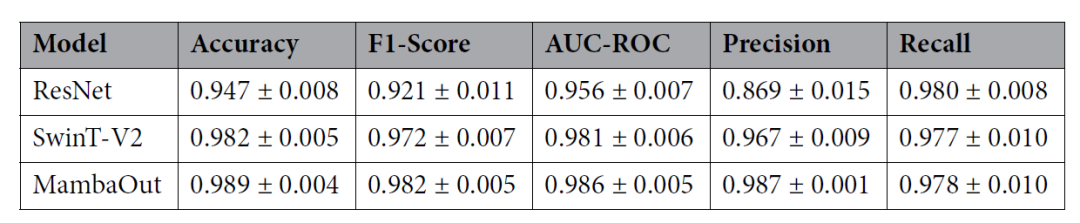
Performance of various deep neural networks on the LyssaScan dataset evaluated at the image level using five-fold cross-validation (mean ± standard deviation)
Finally, since each well corresponds to four images captured from different corner positions, researchers further aggregated the prediction results from image regions to the well level and compared them with the corresponding well-level ground truth annotations to assess whole-well detection performance. The results are shown in the table below:
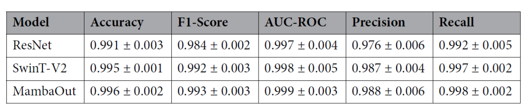
Performance of various deep neural networks on the LyssaScan dataset evaluated at the well level using five-fold cross-validation (mean ± standard deviation)
Across the three evaluation levels—image regions, individual images, and wells—MambaOut consistently achieved the best performance, followed by Swin Transformer V2 (SwinT-V2). At the image region level, MambaOut demonstrated the highest performance in metrics such as accuracy, F1 score, and precision, indicating its ability to reliably detect infection signals within local image regions. This trend was also observed at the image level: MambaOut achieved high accuracy and F1 scores, while SwinT-V2 performed slightly lower than MambaOut.
ResNet exhibited the lowest image-level performance, with an accuracy of 0.947, primarily because this model is sensitive to classification errors at the image region level. Nevertheless, this aggregation rule aligns with practical biological diagnostic logic: the presence of even a single fluorescent positive region indicates the detection of viral antigens, thereby classifying the entire image as infection-positive. In diagnostic scenarios, identifying any evidence of infection is crucial, and false negatives typically have more severe consequences than occasional false positives.
Applicability of Antibody Titer Measurement in Fluorescent Antibody Virus Neutralization (FAVN) Tests
To assess the applicability of the proposed workflow in serological testing scenarios, researchers applied the models to the Fluorescent Antibody Virus Neutralization (FAVN) test. Experimental results showed that MambaOut achieved the best performance, with an accuracy of 0.992, an F1 score of 0.992, and an AUC-ROC of 0.997, indicating that the model can effectively capture local and global features under complex and variable testing conditions. SwinT-V2 also demonstrated strong generalization capabilities, achieving an accuracy of 0.988. In contrast, ResNet showed decreased performance, with an accuracy of 0.965.
Subsequently, researchers further examined its applicability for measuring antibody titers in the FAVN test. Using the same set of images, they compared the antibody titers predicted by the model with those measured by human operators, using the manual interpretation results as the ground truth reference, as shown in the table below:
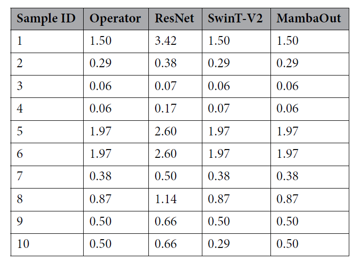
Comparison of antibody titers (International Units per milliliter, IU/mL) predicted by ResNet, SwinT-V2, and Mamba models versus manual operator interpretations for the same sample set
The study employed Bland–Altman analysis to evaluate the agreement between the models and human operators, as shown in the figure below:
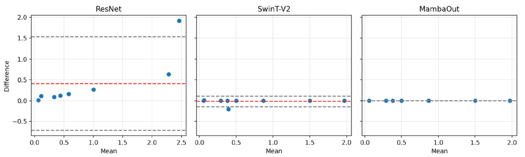
Bland–Altman agreement analysis plot comparing deep learning model predictions with manual operator measurements
ResNet exhibited a significant positive bias, with a mean difference of +0.41 and wide limits of agreement (LoA) ranging from −0.71 to +1.53. This indicates that ResNet tends to systematically overestimate antibody titers compared to manual interpretation results.
SwinT-V2 demonstrated good agreement, with a mean difference close to zero (−0.02) and narrow limits of agreement ranging from −0.15 to +0.11, indicating good interchangeability between its predictions and manual interpretation results across the entire measurement range.
The predictions of MambaOut were perfectly consistent with manual interpretation, with all paired differences being zero.
No evidence of proportional bias was found for SwinT-V2 and MambaOut; however, the differences for ResNet showed a slight increasing trend as antibody titers rose.
According to relevant guidelines from the World Health Organization (WHO) and the World Organisation for Animal Health (WOAH), an antibody titer reaching 0.5 IU/mL can be considered reasonable evidence of seroconversion following vaccination and serves as a reference standard for meeting international pet travel regulations. The study adopted this threshold to evaluate how differences in predicted titers would impact result determination in sample sets covering the full range of antibody titers.
Across the entire sample set, antibody titer estimates produced by ResNet were consistently higher than the actual reference values. This misclassification could result in pets with insufficient immunity being incorrectly cleared for travel.
The overall predictions from SwinT-V2 were relatively close to manual interpretation, but it still misclassified Sample 10 as negative, despite the sample actually meeting the qualification criteria. This could lead to unnecessary re-vaccination and travel delays.
The predictions from MambaOut were perfectly consistent with manual interpretation, correctly identifying all non-qualifying samples and accurately distinguishing between low- and high-titer sera, with no errors in threshold determination.
Taken together, these findings indicate that the proposed system can be effectively applied not only to cell culture detection but also extended to serological testing workflows, supporting its further application in rabies diagnosis and serological surveillance.
Conclusion
In summary, the progress achieved in this study for RABV infection detection further solidifies the scientific foundation for applying computer vision to cell research and diagnostic assays, while also enabling its extension to cell culture detection for other viral pathogens. This study highlights the potential of artificial intelligence to transform laboratory diagnostics, helping to bridge the gap between expert-level interpretation capabilities and scalable, user-friendly technologies. Beyond technical contributions, this approach demonstrates the potential of AI-driven diagnostic technologies to reduce the global burden of rabies. Although the current protocol still relies on specialized equipment, the underlying methodology holds promise for adaptation to more accessible hardware platforms, such as combining standard microscopes with digital cameras, thereby paving the way for scalable and sustainable diagnostic solutions in resource-limited settings.
References:
https://phys.org/news/2026-10-ai-rabies-world.html
https://www.nature.com/articles/s41598-026-57955-8