Command Palette
Search for a command to run...
AI's Breakthrough in Pathology: Coloring Images to Make Them Look Real

By Super Neuro
Scenario description:Using machine learning methods, we can virtually stain the microscopic images of tissue sample slices in pathology to avoid the drawbacks of traditional staining methods and assist medical personnel in making diagnoses more conveniently.
Keywords:CNN, image processing, medical assistanceclose

Medical diagnosis is often done through image observation, and when it comes to image processing, AI is very useful.
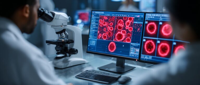
In pathological examinations of biopsy tissue sections, it is necessary to stain extremely thin sections of the sample and then observe the image under a microscope for pathological diagnosis. From the perspective of AI, this problem is a matter of accurately coloring the image.
In a recent report, researchers used machine learning methods to achieve extremely high accuracy in virtual staining of slices, which can basically replace the manual staining process.
Traditional staining of tissue sample sections
Microscopic imaging of tissue samples is a fundamental tool for diagnosing a wide range of diseases and is a workhorse of pathology and the biological sciences.
The specific operation is to remove a very small piece of body tissue and process and analyze the sample to achieve the purpose of examination and diagnosis.

After the sample is removed, it is sliced into thin sections, a few micrometers (millionths of a meter) thick. These thin tissue sections contain information about the patient's condition at a microscopic scale.
Under a standard optical microscope, unprocessed sections are almost indistinguishable. Staining can only be used to increase identification. In the development of pathology over the years, doctors have created many tissue staining methods.
However, the traditional staining process of tissue specimens is time-consuming and complicated, requiring specialized laboratory infrastructure, chemical reagents and well-trained technicians.
Digital coloring with AI
So how does AI perform coloring?
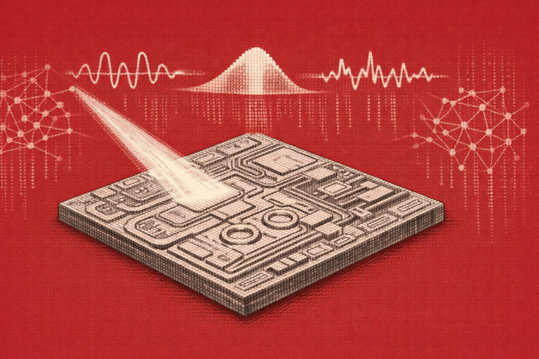
Virtual image staining utilizes a machine learning approach by using a deep convolutional neural network (CNN) to colorize a single autofluorescence image of a sample using data from previous stainings.
During the operation, first slice the unstained tissue and take a microscopic image of it under autofluorescence.
Then, using a CNN trained with a generative adversarial network (GAN), the unlabeled tissue autofluorescence image can be quickly converted into an image similar to reagent staining.

This deep learning-based method does not involve the tedious traditional steps at all. It trains the model through a computer and finally outputs a colored image, which can greatly save costs and time.
The research was conducted by a research team from the University of California, Los Angeles, and the results were published in the journal Nature Biomedical Engineering.
Paper address: https://www.nature.com/articles/s41551-019-0362-y
Is it really that good?
So what is the actual effect of AI virtual coloring?
To judge the effectiveness of virtual dyeing, the researchers used a "blind review" process.
Board-certified pathologists were asked to make independent judgments without being informed whether the samples were stained with reagents or with AI virtual staining.
The final conclusion shows that in terms of staining quality, the medical diagnosis produced by AI-generated virtual staining has no clinically significant difference compared to previous methods.
The researchers also stained some samples using conventional methods after virtual staining, and the resulting images showed that the actual effects were almost the same.

The first column is the contrast-enhanced image, the second column is the original autofluorescence image, the third column is the AI virtual staining, and the fourth column is a traditional method: Masson trichrome staining. The samples are liver and lung slices.
The new method was validated with different stains and human tissue types, including routine sections of salivary glands, thyroid, kidney, liver, and lung.
They said that the next step is to conduct large-scale randomized clinical studies to verify the accuracy of AI staining image diagnosis.
What technology can do
The AI staining method only requires a standard fluorescence microscope and a simple computer, so it has transformative advantages in resource-limited environments and conditions.
"This technology has the potential to transform clinical histopathology workflows," said Aydogan Ozcan, who led the study. "Because the technology is accessible, the staining process becomes fast and simple, and does not require specialized technicians or advanced medical laboratories."
Regarding the scalability of this method, he added, "The AI-based virtual staining framework can also be used in the operating room, for example, to quickly assess tumor margins and provide convenient or even critical guidance to surgeons performing surgery."
In addition, another major impact of this study is that it helps standardize the entire staining process. The use of AI methods can prevent differences caused by different technicians and operating environments, thereby avoiding misdiagnosis or misclassification of biopsies.

